Knee Pain | Cartilage Injuries
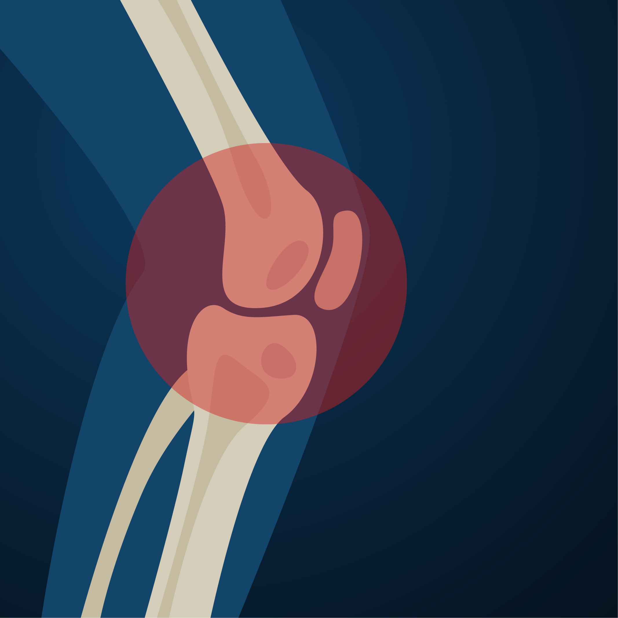
Knee pain is a common cause of disability for patients of all ages. The knee is a complex joint that is made up of three different bones – the femur, the tibia, and the patella. The knee is held together with complex arrangements of ligaments that create a modified hinge joint.
Cartilage is the material that covers the ends of the bones where they contact to become the knee joint. Articular cartilage is a very durable material that provides a smooth and impact-resistant surface and allows the bones to glide over one another during the knee range of motion required during activities such as walking, running, and jumping.
Many factors can contribute to cartilage damage/injuries including age-related degeneration of cartilage, excessive weight, repetitive or acute injuries. During normal activities of life, the cartilage is exposed to extreme pressure. This pressure is increased significantly during activities such as running and jumping. It can be injured from this repetitive stress or from a single traumatic event. The cartilage surface can be sheared off the end of the bone, exposing the underlying bony surface. Unlike other tissues in our body, injured cartilage does not have the ability to heal.
Cartilage lesions left untreated may by themselves cause pain and loss of functions. Further, they may progress and lead to early degenerative arthritis. Once full-blown arthritis is present, symptoms become worse and are often debilitating.
Diagnosing a cartilage injury can be difficult as the symptoms can vary greatly and the onset of symptoms can occur suddenly or come on very slowly. The symptoms of cartilage injuries include pain with weight-bearing, catching or locking, giving way or buckling, and swelling.
Additionally, injuries to cartilage are often difficult to diagnose by physical examination. Diagnostic imaging such as X-rays and MRIs can help but are also not perfect in their ability to diagnose these lesions. It is often necessary to perform a direct inspection of the joint via arthroscopy to diagnose this problem.
Arthroscopy is a minimally invasive surgical procedure where the surgeon inspects the joint with a camera. With this camera, the surgeon can inspect the joint surfaces and determine the size, location, and severity of the injury. All of these factors will determine the best treatment option for the injury. Sometimes the surgeon will immediately treat the injured cartilage; other times, the surgeon may determine that additional surgery is necessary to deliver the best care.
There are several treatment options for articular cartilage lesions:
Chondroplasty
This is performed during the initial arthroscopic procedure and is where any loose cartilage is cleaned out (detrided) from the joint. The surgeon attempts to create a “stable” lesion by removing any loose flaps or pieces of cartilage that are loose. Chondroplasty can help alleviate symptoms, but it does not actually repair the lesion.
Microfracture
This procedure is also performed arthroscopically during the initial procedure and is an attempt to get the body’s own marrow cells to initiate a healing response. Unfortunately, the healing response does not create normal cartilage, but a form of scar tissue that is not as durable as normal articular cartilage.
This procedure is generally used for smaller lesions. During the procedure, a special awl or small drill bit is used to create tiny holes in the bone to access the bone marrow.
With these access holes, the marrow cells seep into the defect and initiate a “healing response”.
Osteochondral Autrograft
This is also performed during the initial arthroscopic procedure and can be performed arthroscopically or through formal open incisions. It is where a “donor” plug of healthy bone and cartilage is taken from an area of the knee that is not weight-bearing and placed into the affected area. This procedure is typically utilized for only small defects.
Osteochondral Allograft
This procedure is very similar to the autograft procedure but because of the size of the lesion, you cannot take a large enough “donor plug” from the patient’s knee without creating significant morbidity. Therefore, this procedure is done utilizing fresh cadaver bone and cartilage that has been size-matched to the patient.
Autologous Chondrocyte Implantation (ACI)
ACI utilizes the patient’s own cells to repair the cartilage lesion. To perform an ACI, the surgeon takes a biopsy of the patient’s healthy cartilage during the initial arthroscopy. The cartilage is sent to Genzyme’s lab where they culture the cartilage cells and grow them in the laboratory to create millions of new and healthy cartilage cells. During a second surgery, the surgeon implants these cells back into the injured area where they form new, healthy articular cartilage.
"*" indicates required fields
Our Location
Dr. Christopher K. Jones, MD
4110 Briargate Parkway #300
Colorado Springs, Colorado 80920
Hours
Monday: 9am-5pm
Thursday 9am-5pm
Friday 9am-5pm

