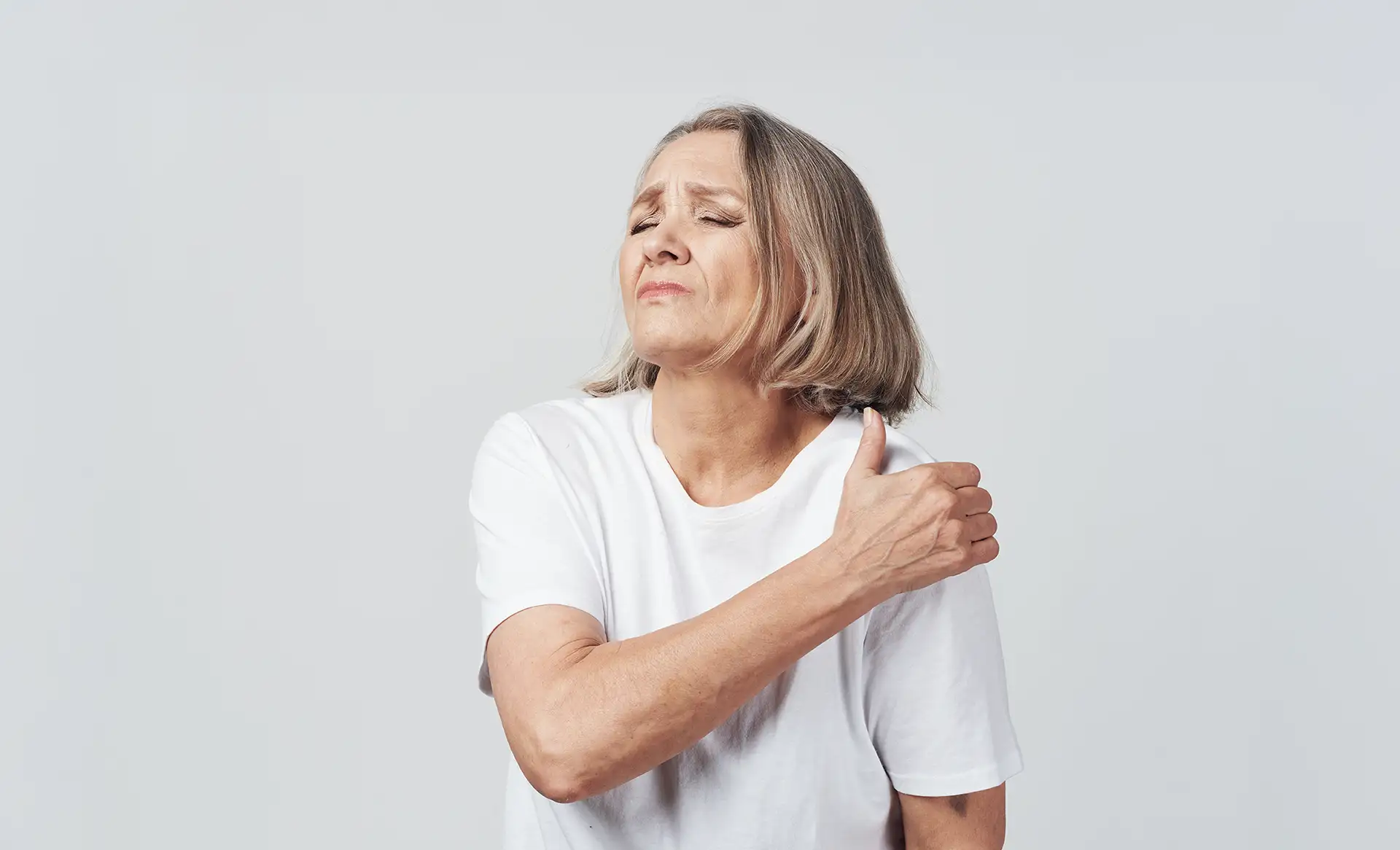
The shoulder joint offers the greatest range of motion of any joint in the body. This incredible mobility, however, comes at the cost of inherent instability, making it susceptible to a wide array of injuries and degenerative conditions. For active individuals, athletes, and those who rely on their shoulders for daily life, a debilitating shoulder injury can be a profound source of frustration and pain. When non-surgical treatments fail to provide lasting relief, surgery may become a recommended option.
Arthroscopic shoulder surgery is a minimally invasive surgical technique, offering many patients a potential path to reduced pain and a return to activities. In this post, we’ll explore arthroscopic shoulder surgery in detail, empowering you to make the most informed decision about your orthopedic health.
Key Takeaways
- Arthroscopy uses a small camera (arthroscope) and miniature instruments inserted through tiny incisions (portals), which may result in less soft tissue damage and a quicker initial recovery compared to traditional open surgery in many cases.
- This technique may be used to treat numerous conditions, including rotator cuff tears, shoulder instability (dislocations), labral tears (SLAP and Bankart lesions), shoulder impingement, and frozen shoulder.
- While the surgery is minimally invasive, the success of the procedure is typically dependent on a dedicated, structured rehabilitation program.
Understanding the Shoulder Joint
The shoulder is not a single joint but a complex of several joints, muscles, tendons, and ligaments working in concert. It is comprised of three bones: the humerus (upper arm bone), the scapula (shoulder blade), and the clavicle (collarbone).
The primary joint is the glenohumeral joint, a ball-and-socket mechanism where the rounded head of the humerus (the ball) fits into the shallow socket of the scapula, known as the glenoid. This design allows for remarkable rotation and elevation. However, the glenoid socket is quite shallow, meaning the joint relies heavily on surrounding soft tissues for stability, including the labrum, rotator cuff, shoulder capsule, and bursa.
The delicate balance between the shoulder’s extreme mobility and its reliance on these soft tissues for stability explains why it is so vulnerable to injury, particularly in sports and activities that involve repetitive overhead motion.
What is Arthroscopic Shoulder Surgery?
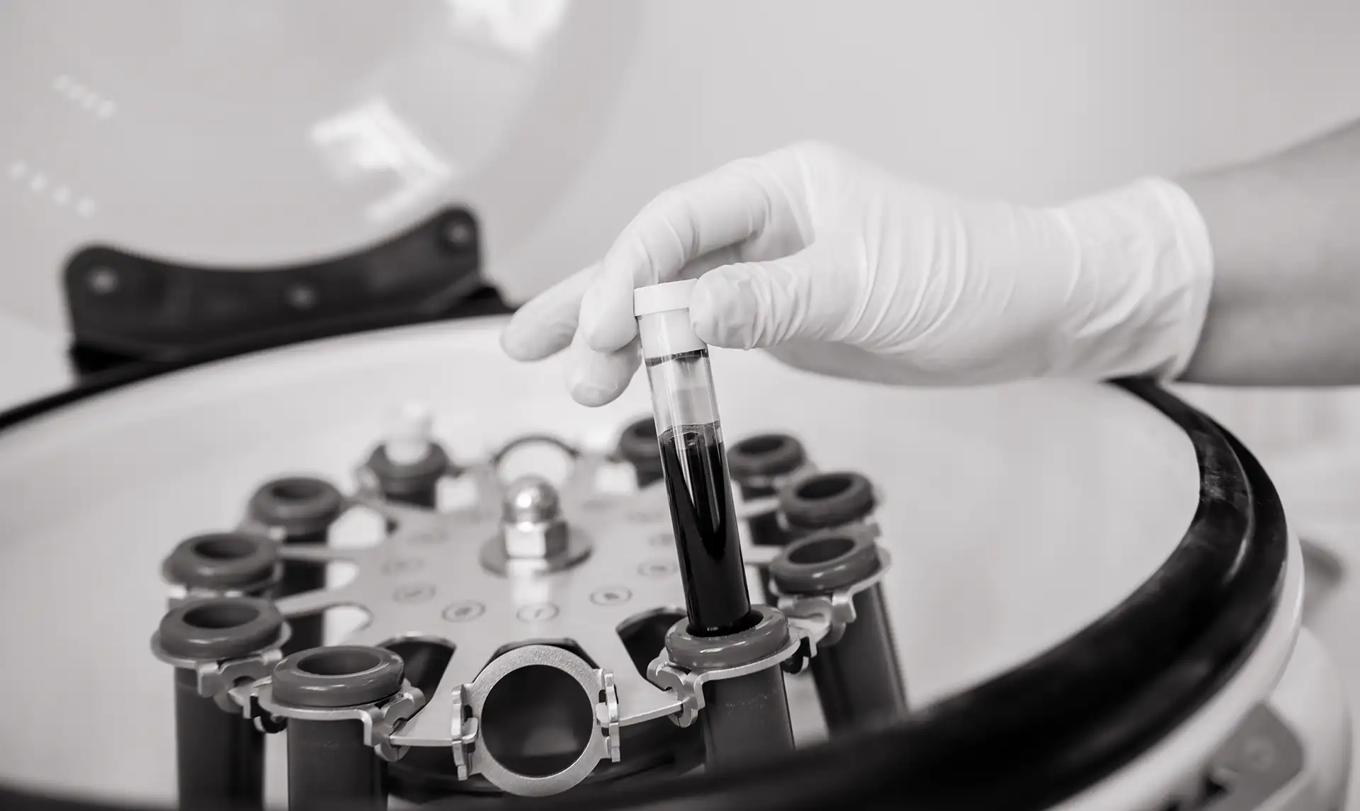
Shoulder arthroscopy is a surgical technique that allows orthopedic surgeons to view, diagnose, and treat problems inside a joint without making a large incision. Instead of fully opening the joint, the surgeon makes several small incisions, typically less than a centimeter long, called portals.
Through one portal, the surgeon inserts an arthroscope, a pencil-sized instrument containing a lens, a light source, and a small video camera. This camera transmits real-time, magnified images of the joint’s interior onto a high-definition video monitor.
Through the other portals, the surgeon inserts specialized, miniature surgical instruments designed to perform precise tasks, such as cutting, shaving, grasping, and suturing. The joint is continuously irrigated with a sterile fluid to expand the space and wash away debris, ensuring a clear field of view.
When is Arthroscopy Recommended? Indications for Surgery
Arthroscopic shoulder surgery is frequently recommended when a painful condition does not respond to a comprehensive course of non-surgical treatment. The procedure is versatile and can be used to treat a wide spectrum of shoulder pathologies, some of which we’ll explore in the following subsections.
Rotator Cuff Tears
Rotator cuff tears can be partial (fraying of the tendon) or full-thickness (the tendon is completely separated from the bone). Your surgeon may use arthroscopy to reattach the torn tendon to the head of the humerus using specialized anchors and high-strength sutures. The goal of the repair is to restore the integrity of the tendon-to-bone attachment. The success of a repair can be dependent on the quality of the tissue, the size of the tear, and the patient’s commitment to the post-operative rehabilitation protocol.
Shoulder Instability and Labral Tears
Shoulder instability occurs when the head of the humerus is forced out of the glenoid socket, either partially (subluxation) or completely (dislocation). This may result in damage to the labrum and the ligaments.
- Bankart Repair: A Bankart lesion is a tear of the lower part of the labrum, which occurs when the shoulder dislocates forward. Arthroscopic Bankart repair may involve reattaching the torn labrum and tightening the stretched ligaments to the bone, restoring the shoulder’s stability.
- SLAP Repair: A SLAP tear involves the upper part of the labrum, often extending into the biceps tendon attachment. These tears are common in overhead athletes (e.g., baseball pitchers, tennis players). The arthroscopic procedure typically involves either repairing the torn labrum or, in older patients, performing a biceps tenodesis (releasing the biceps tendon from the labrum and reattaching it to the humerus bone) to eliminate the source of pain.
Shoulder Impingement Syndrome (Subacromial Decompression)
Impingement occurs when the rotator cuff tendons are pinched between the head of the humerus and the acromion (the bone on top of the shoulder) during arm elevation. This pinching can be caused by a bone spur on the underside of the acromion or inflammation of the bursa. Arthroscopic subacromial decompression involves removing the inflamed bursa and shaving away the bone spur from the underside of the acromion. This procedure creates more space for the rotator cuff tendons to glide freely.
Frozen Shoulder (Adhesive Capsulitis)
Frozen shoulder is a condition characterized by severe stiffness and pain, caused by the thickening and tightening of the shoulder capsule. While often treated non-surgically, if symptoms are severe and persistent, arthroscopy may be used to perform a capsular release. During this procedure, the surgeon uses small instruments to carefully cut and release the tight, contracted portions of the joint capsule, with the goal of restoring a significant amount of motion.
Other Arthroscopic Procedures
Arthroscopy may also be used for many other procedures, including removal of loose bodies (fragments of cartilage or bone within the joint), treatment of arthritis-related inflammation, repair of small cartilage defects, and debridement (cleaning out frayed or damaged tissue) to smooth the joint surfaces and reduce irritation.
The Arthroscopic Procedure
Understanding the steps involved in the surgery can help prepare you for the day of the procedure. Arthroscopic shoulder surgery is typically performed as an outpatient procedure, meaning you can go home the same day.
Pre-Operative Planning and Preparation
Before the surgery, your orthopedic surgeon will conduct a thorough evaluation to ensure you are medically fit for the procedure. You will also receive specific instructions regarding:
- Medications: You may need to stop taking certain medications for a period before surgery.
- Fasting: You should follow strict instructions regarding when to stop eating or drinking prior to the procedure, typically after midnight the night before.
- Logistics: Arranging for a responsible adult to drive you home and stay with you for at least the first 24 hours is important.
Anesthesia
A member of the anesthesia team will discuss your options, which often involve a combination of general anesthesia and a regional nerve block:
- General Anesthesia: You will likely be completely asleep for the duration of the surgery.
- Regional Anesthesia (Interscalene Block): This involves injecting a local anesthetic around the nerves that supply the shoulder and arm. This block provides pain relief for the first 12 to 24 hours after surgery, reducing the need for strong pain medication immediately post-op and making the initial recovery phase more comfortable.
Positioning and Incisions
Once the anesthesia is administered, the surgical team will position you for the procedure. The surgeon then makes the small, precise portals. These incisions are strategically placed to allow the surgeon to access all necessary areas of the joint while minimizing damage to the surrounding soft tissues.
Visualization and Repair
- Fluid Infusion: Sterile fluid is pumped into the joint to gently inflate the space, which improves visibility and helps control bleeding.
- Diagnosis: The arthroscope is inserted, and the surgeon performs a systematic inspection of the entire joint to confirm the diagnosis and assess the full extent of the damage.
- The Repair: Using the images on the monitor as a guide, the surgeon inserts the specialized instruments through the other portals to perform the necessary repair.
- Closure: Once the repair is complete, the instruments and arthroscope are removed, the fluid is drained, and the small portals are closed with one or two stitches or sterile adhesive strips.
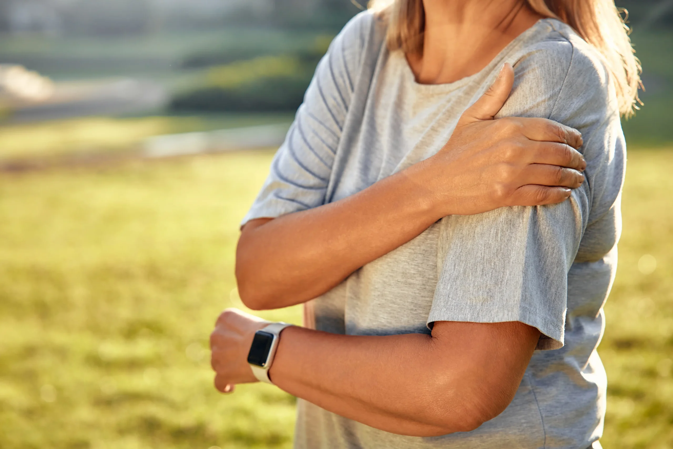
Recovery and Rehabilitation
A key component of a successful outcome is the post-operative recovery and rehabilitation program. Immediately following the procedure, you will spend time in the recovery room as the anesthesia wears off. Before discharge, the nursing staff will ensure your pain is controlled and you meet all discharge criteria. Your arm will likely be in a sling or immobilizer. The duration and type of immobilization depend on the procedure performed.
Rehabilitation after arthroscopic shoulder surgery is a gradual process designed to restore motion, strength, and function while protecting the repair. Though each patient’s recovery plan is individualized, it generally follows four key phases.
Phase I (Protection): The initial weeks focus on rest, reducing pain and swelling, and protecting the surgical repair. Only gentle, passive movements, often assisted by a physical therapist, are performed.
Phase II (Early Strengthening): As healing progresses, patients may begin moving the arm actively and performing light strengthening and stretching exercises to restore range of motion.
Phase III (Advanced Strengthening): Resistance training and targeted exercises for the rotator cuff and shoulder blade muscles can help rebuild strength and stability.
Phase IV (Return to Activity): The final stage emphasizes regaining function and safely returning to sports, work, or daily activities.
Summary
Arthroscopic shoulder surgery is a testament to the advancements in orthopedic medicine, offering a minimally invasive solution for a wide range of painful and debilitating shoulder conditions. From repairing a torn rotator cuff to stabilizing a chronically dislocating joint, this technique may help restore the complex mechanics of the shoulder while minimizing the disruption to your life.
Frequently Asked Questions
How long does arthroscopic shoulder surgery typically take?
The duration of the surgery depends on the complexity of the repair. A simple procedure, such as a subacromial decompression, may take less than an hour. A complex repair involving a large rotator cuff tear or multi-ligament stabilization may take between 90 minutes and two hours. Your surgeon will provide a more accurate estimate based on your specific condition.
Will I have to stay overnight in the hospital?
In the vast majority of cases, arthroscopic shoulder surgery is performed as an outpatient procedure. This means you will arrive at the surgical center or hospital, have the surgery, spend a few hours in the recovery room, and be discharged home the same day. You must have a responsible adult available to drive you home and assist you for the first night.
Will I need physical therapy, and how long will it last?
Oftentimes, physical therapy is important for an optimal outcome. The total duration of therapy typically lasts around three to six months, though the frequency of visits may decrease over time as you progress to a home exercise program.
How long until I can return to sports or overhead activities?
Returning to sports is a gradual process guided by your surgeon and physical therapist, and timelines may significantly vary depending on the patient and the procedure. For many patients, a return to light, non-contact sports may begin around three to four months. A full return to contact sports, throwing, or heavy overhead activities is typically not permitted until six months or later post-surgery, and only after passing specific strength and functional tests.